All About Anatomy
- Gillian Higgins
- Oct 23, 2023
- 4 min read
Updated: Jun 5, 2024
The Horses Inside Out Anatomy Exhibition is a must see for every equestrian and anatomy enthusiast and one to rival aspects of the Natural History Museum! Each skeleton, specimen and anatomical model reveals different fascinating structure, anatomy, asymmetries, anomalies and pathologies.
The next anatomy exhibition will take place at the 2024 Conference: Growth and Development. There is a great opportunity to get up, close and personal with each specimen, get hands on with the interactive models and ask the inevitable ‘Why-How-What..? questions that anatomy conjures.
To whet your appetite for all things anatomical, I thought I’d give you a little overview of what will be coming to the anatomy exhibition in February 2024 (including some exciting new models). You’ll have to come along and see for yourself which ones we will have put on display! Follow this link to find out more: https://www.horsesinsideout.com/c24
First up we have a number of skeletons demonstrating a range of ages and poses. This gives a great opportunity to compare and contrast: Growth and Development - A Journey of Lifetime. All are brilliant examples of their age and size and being able to compare very young, young, middle age and older, well-used bones is very interesting. Don't forget to look out for the growth plates.
Yearling Skeletons
We have 2 immature skeletons and it is fascinating to see all the growth plates still open. Epiphyses (growth plates) are a type of joint, a cartilaginous joint, which ossify when the bone finishes growing. The age at which the growth plates fuse tend to follow a pattern and once all the growth plates have fused the skeleton is mature.
A Four Year Old Skeleton
The skeleton of a 4yo 15.1hh Welsh Section D assembled in a cantering pose is particularly fascinating as it is partially immature and shows which growth plates are still open at the age at which most horses are backed and started being ridden. There are also some developmental anomalies.
Middle Age Skeleton
The skeleton of a 11yo 13.2hh Dartmoor pony in standing position shows a mature bones with few problems.
Veteran Skeleton
Freddie Fox, my heart horse, much-loved eventer and original star of Horses Inside Out a 16.2hh 24yo skeleton assembled in a trotting pose. As an older skeleton there are a number of bony changes and signs of wear and life.
Anatomical models
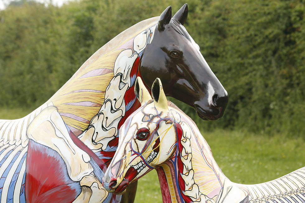
Meet Leonardo, named such after Leonardo Da Vinci, a model foal painted up with the superficial muscles on one side and the skeleton including the growth plates on the other.
Leonardo is a work of art and a great example of commissioned models Gillian creates.
Tendons and Ligaments of the Lower Limb
The air-dried equine forelimb is one of my favourite specimens as it beautifully illustrates the tendons and ligaments. The method used to preserve this leg means the tendons and ligaments still remain attached to their bony landmarks albeit, dehydrated and contracted.
If ever you have had trouble trying to remember which ligament lies closest to the bone and which lies further away, check this limb out. And talking of checks, you can even see the check ligaments, too!
This is the leg that I used in the online seminar 'Understanding Tendons and Ligaments of the Lower Limb' to illustrate the anatomy, biomechanics and palpation.
Anatomical Head Models
We have a number of skulls and anatomical head models. Look out for all the holes that allow for the nerves to pass from the inside of the head to the outer structures of the face. And don’t forget to look for the hyoid bones. If you can name all the different parts that make up the whole of the hyoid system, I’ll buy you a coffee!
I am particularly proud of Posideon, a 3D printed model of a horse’s head. I originally sculpted the muscles, nerves and blood vessels out of plasticine before scanning it, printing and then painting. One of my favourite aspects of this head model is how it shows the hyoid bones and the connection into the horse's tongue.
Poseidon will be instantly recognisable if any of you have bought the beautiful book Illustrated Head Anatomy This comprehensive book illustrating all the external structures of the head with models, painted horses and dissection photographs is must read for all bridle fitters and equine dentists - in fact, anyone interested in the anatomy of the head and it's biomechanical connections.
The Carpus and Tarsus
The carpus, commonly called the knee but equivalent to our wrist and the tarsus, the hock, are the 2 of the most complicated joints in the horse and they are some of the hardest bones to assemble. With the 2 magnetic models that repel bones when the bones are in the incorrect place and magnetically link together all those little bones in the correct order. Have a go yourself, time your efforts and see if you can beat your friend
Anatomy is truly amazing stuff and our Anatomical Exhibition gives you a fantastic opportunity to not only look at the goings on inside a horse, but also to compare structures from one horse to another. If this article has whetted your appetite, do come along and have a good wander around our display. I promise there will be so much more than illustrated in this article and you won't be disappointed!
We are looking forward to seeing you there!
Gillian xx
To purchase recordings of the conference seminars, follow this link: www.horsesinsideout.com/c24


I loved the anatomy gallery at last year's conference and learned a lot. This year at the conference I expect to learn a lot more Journey of my horse's Lifetime is an intriguing title and I know I'll appreciate and use what I learn.
Thanks so much for sharing all that you do. You are helping all of us do the best we can to kheep our horses sound and performing well.